What Your Dentist Learns From Comparing Current And Previous Dental Images
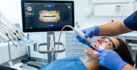
You sit back in the chair, the images come up on the screen, and your dentist starts looking at today’s X rays next to ones from years ago. If you have ever wondered what they are actually looking for, you are not alone. A lot of people hear “we’re comparing your films” and assume it is routine, but the comparison can reveal changes that are easy to miss in a single snapshot. Even patients searching for Best clear aligners in Dearborn Heights can benefit from understanding how these image comparisons help dentists spot subtle dental changes over time.
That matters because teeth and bone do not usually change all at once. Problems often build slowly. A small dark spot between teeth, a slight drop in bone height, a filling that no longer seals the edge of a tooth, these things can stay quiet for a long time. When your dentist compares old and new images, they are not just checking for cavities. They are tracking movement, loss, healing, wear, and patterns that tell a bigger story about your oral health. Comparing dental X rays over time helps your general dentist decide whether to watch, treat, or reassure you.
Comparing dental images shows change, and change is what guides treatment
A single dental image shows what is there today. A series of images shows what is changing. That difference shapes care.
Your dentist may compare bitewing, panoramic, or periapical images to look for new decay between teeth, bone loss from gum disease, infection around a root, shifting tooth position, or changes under crowns and fillings. According to the American Dental Association’s overview of dental radiographs, dental X rays help detect conditions that may not be visible during a visual exam alone.
That comparison becomes even more useful when nothing hurts. Early decay often has no symptoms. Bone loss from periodontal disease can progress quietly. A tooth with a past root canal may look stable for years, then show a small change at the tip of the root that signals inflammation or infection. If your dentist only saw the current image, the finding might look minor or uncertain. Side by side with an older image, the pattern becomes clearer.
This is also where stress creeps in for patients. You may hear that something is “different from last time” and worry that it means a major problem or a major bill. Sometimes it does mean treatment is needed. Sometimes it means the opposite. A spot that has not changed in five years may be watched instead of drilled. Stable bone levels may confirm that your home care and gum treatment are working. Comparison protects you from under-treatment and over-treatment.
Past and present dental radiographs reveal disease, healing, and wear
Current and previous dental images give your dentist a timeline. That timeline can answer practical questions that a mirror and explorer cannot.
If a filling was placed a few years ago and the new image shows a shadow at the edge, your dentist checks the older image to see whether that shadow is new, larger, or unchanged. If you grind your teeth, images can show how pressure is affecting bone support or restorations over time. If you had an extraction or implant, comparison helps assess healing and bone response.
Research has also shown the value of comparing radiographs to improve diagnosis and treatment planning. One study available through PubMed Central on dental radiographic interpretation discusses how image review supports the detection of pathology and structural change. Another source on radiographic assessment in dental care reinforces that serial imaging can help clinicians monitor progression and outcomes over time.
This is one reason your records matter. If you move, switch offices, or skip regular visits for a few years, the lack of prior images can make decision-making harder. Your dentist can still evaluate the current condition, but they lose the ability to measure pace. Is that area of bone loss old and stable, or recent and active? Is that calcification harmless and unchanged, or new and worth more attention? Comparing dates often answers the question.
What your general dentist can learn from image comparisons
| What is being compared | What your dentist may learn | What it can mean for you |
|---|---|---|
| Bone levels around teeth | Whether gum disease is stable or progressing | Home care changes, deep cleaning, periodontal follow up, or reassurance |
| Dark areas between teeth | Whether decay is new, growing, or unchanged | Monitoring, fluoride support, or a filling |
| Edges of crowns and fillings | Whether a restoration is leaking, breaking down, or still sealed | Repair, replacement, or no treatment yet |
| Root tips and surrounding bone | Whether infection, inflammation, or healing is present | Root canal evaluation, retreatment, referral, or continued observation |
| Impacted or shifting teeth | Whether teeth are moving or affecting nearby structures | Orthodontic review, extraction planning, or monitoring |
This is the practical value of dental image comparison. It gives context. Context leads to better timing, and better timing often means simpler treatment.
Three steps help you get more from your dental imaging
Ask what changed, not just what was found. If your dentist points out an area on your X ray, ask whether it is new, bigger, smaller, or stable. That one question gives you a clearer sense of urgency.
Keep your records moving with you. If you change dental offices, request copies of prior radiographs. Older images can save time, reduce repeat work, and help your new provider make more accurate decisions.
Stay consistent with routine visits. The longer the gap between exams, the harder it is to catch slow changes early. Regular imaging based on your risk level helps your general dentist spot patterns before they turn into pain or expensive repair.
Comparing dental X rays helps your dentist treat you with more precision
When your dentist studies old and new images together, they are looking for a story your mouth has been telling over time. Sometimes that story says a problem is growing and needs care. Sometimes it says the area is stable and can be watched. Both answers matter.
If you have been told your dentist wants to compare radiographs, that is usually a sign of careful care, not alarm. It means they want evidence before making a recommendation. That kind of attention can spare you from guesswork and help protect your teeth, bone, and budget.
Bring your questions to your next appointment and ask your dentist to show you the changes on the screen. Seeing the difference for yourself often makes the plan easier to understand and easier to trust.